
A fontanelle, also known as a “soft spot”, is an anatomical feature of the infant human skull comprising any of the soft membranous gaps (sutures) between the cranial bones that make up the calvaria of a fetus or an infant. Fontanelles allow for rapid stretching and deformation of the neurocranium as the brain expands faster than the surrounding bone can grow. Premature complete ossification of the sutures is called craniosynostosis.
During infancy, the anterior fontanelle is known as the bregma.
The skull of a baby consists of five main bones: two frontal bones, two parietal bones, and one occipital bone. These are joined by fibrous sutures, which allow movement that facilitates childbirth and brain growth.
Posterior fontanelle is triangle-shaped. It lies at the junction between the sagittal suture and lambdoid suture. At birth, the skull features a small posterior fontanelle with an open area covered by a tough membrane, where the two parietal bones adjoin the occipital bone (at the lambda). The posterior fontanelles ossify within 2 or 3 months of birth. This is called intramembranous ossification. The mesenchymal connective tissue turns into bone tissue.
Anterior fontanelle is a diamond-shaped membrane-filled space located between the two frontal and two parietal bones of the developing fetal skull. It persists until approximately 18 months after birth. It is at the junction of the coronal suture and sagittal suture. The fetal anterior fontanelle may be palpated until 18 months. In cleidocranial dysostosis, however, it is often late in closing or may never close. Examination of an infant includes palpating the anterior fontanelle.
Two smaller fontanelles are located on each side of the head, more anteriorly the sphenoidal or anterolateral fontanelle (between the sphenoid, parietal, temporal, and frontal bones) and more posteriorly the mastoid or posterolateral fontanelle (between the temporal, occipital, and parietal bones).
During birth, fontanelles enable the bony plates of the skull to flex, allowing the child’s head to pass through the birth canal. The ossification of the bones of the skull causes the anterior fontanelle to close over by 9 to 18 months. The sphenoidal and posterior fontanelles close during the first few months of life. The closures eventually form the sutures of the neurocranium. Other than the anterior and posterior fontanelles, the mastoid fontanelle and the sphenoidal fontanelle are also significant.
The sequence of fontanelle closure is as follows:
The posterior fontanelle generally closes 2 to 3 months after birth;
The sphenoidal fontanelle is the next to close around 6 months after birth;
The mastoid fontanelle closes next from 6 to 18 months after birth; and
The anterior fontanelle is generally the last to close between 18-24 months.
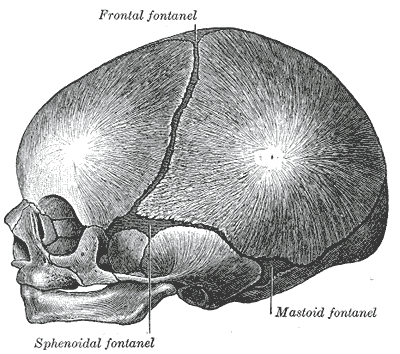
Infant skull at birth, showing the lateral fontanelles.

The anterior and posterior fontanelles.
Drawings courtesy of Gray’s Anatomy (Public Domain)
Fontanelle. (2016, October 4). In Wikipedia, The Free Encyclopedia. Retrieved 03:15, October 4, 2016, from https://en.wikipedia.org/w/index.php?title=Fontanelle&oldid=742505169